


The 13th IASTED International Conference on
Biomedical Engineering
BioMed 2017
February 20 – 21, 2017
Innsbruck, Austria
TUTORIAL SESSION
Medical image analysis using deep learning
Duration
3 hours
Abstract
Medical image analysis is an active and vibrant research area, which has become more relevant
during the past two decades, thanks to developments and advances in multidimensional
medical imaging modalities, such as X-ray computer tomography (X-ray CT), ultrasound, positron emission tomography (PET), magnetic resonance imaging (MRI) and functional MRI (fMRI), among others. The development of more advanced imaging instrumentation triggered
comparable scientific advances in the field of computer-based medical image processing, for
tasks such as image reconstruction, image processing, and – of central interest for this tutorial –
image analysis techniques that can lead to better understanding and interpretation of medical
images.
In parallel with the advances in medical imaging, the field of artificial intelligence (AI) –
particularly machine learning – has also experienced significant growth during the past 20 years,
and many intelligent systems for medical diagnosis have been developed both in industry and
academia. More recently, the emergence of a machine learning paradigm known as deep
learning has further energized the field and enabled the development of medical image analysis
systems that can display remarkable accuracy, to the point of raising concerns about the future
of the human radiologist.
Combined, these developments have resulted in an unparalleled and unprecedented state of
affairs, where many challenging research problems are being solved and new market
opportunities have started to emerge.
In this tutorial we provide an extensive overview of the field of medical image analysis, with
emphasis on the recent impact of increasingly popular deep learning techniques on the design
and implementation of intelligent medical imaging-based diagnosis systems.
Objectives
At the end of this tutorial, participants should be able to:
• Understand the fundamentals of medical image analysis techniques.
• Appreciate the challenges involved in designing medical image analysis systems using
machine learning techniques.
• Assess the impact of deep learning networks, algorithms, techniques, and frameworks in
the field of medical image analysis.
• Identify directions for potential research in this field.
Timeline
1. Motivation: "Will Computers Replace Radiologists?" (10 min)
2. Fundamentals of Medical Image Analysis (40 min)
a. Medical imaging modalities
b. Medical image processing and enhancement
c. Medical image representation, analysis, and classification
d. Medical image retrieval
e. Selected tools for scientists and developers
3. Fundamentals of Artificial Intelligence (AI) and Machine Learning (ML) (40 min)
a. Brief history of AI
b. Foundational concepts
c. Popular ML techniques
d. Practical hints and best practices
e. Selected tools for scientists and developers
4. Designing medical image analysis systems using ML techniques (20 min)
a. Problem definition
b. Challenges
c. Examples, case studies and success stories
5. Deep Learning (30 min)
a. The deep learning phenomenon
b. Basics of neural networks
c. Fundamentals of deep learning techniques and algorithms
d. Deep learning frameworks
e. Examples of successful deep learning applications and results
6. Deep Learning in medical applications (30 min)
a. Image description using deep learning techniques
b. Medical image reconstruction using deep learning
c. Medical imaging-based diagnosis using deep learning
d. Examples of deep learning-oriented applications in the medical imaging domain
e. Datasets, challenges, and benchmarks
f. Our work (skin lesion segmentation and classification using deep learning)
7. Reflection (10 min)
a. Is deep learning overrated?
B. Will computers replace radiologists?
C. What are the most promising directions for research in this field?
Target Audience
researchers and practitioners with familiarity with (medical) image processing and analysis and/or machine learning.
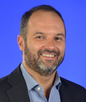
Qualifications of the Instructor(s)
Dr. Oge Marques is Professor of Engineering and Computer Science at Florida Atlantic University. He is Tau Beta Pi Eminent Engineer, ACM Distinguished Speaker, and the author of more than 100 publications in the area of intelligent processing of visual information. Professor Marques is Senior Member of both the IEEE and the ACM and member of the honor societies of Sigma Xi, Phi Kappa Phi and Upsilon Pi Epsilon. He has more than 30 years of teaching and research experience in different countries (USA, Austria, Brazil, India, Spain, France, and the Netherlands).